
Shoulder MRI vs Ultrasound Jaipur
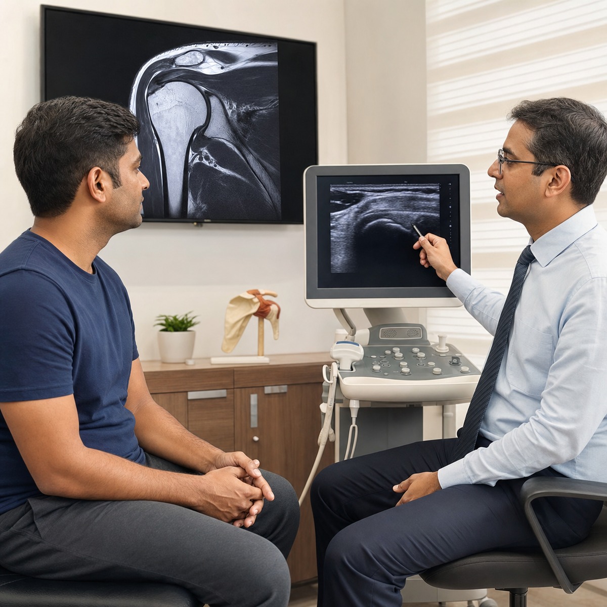
Quick answer: Shoulder MRI vs ultrasound Jaipur is not a contest with one winner for every patient. Ultrasound can assess rotator cuff tendons dynamically and is often accessible, but its accuracy depends on the operator. MRI gives a wider view of tendons, muscles, cartilage and bone marrow, making it useful for complex injuries and surgical planning.
What matters for this shoulder decision
- A clinical examination should define the question before any shoulder scan is ordered.
- Ultrasound is strong for dynamic rotator cuff and biceps tendon assessment in experienced hands.
- MRI shows deeper structures and muscle quality and is often preferred for complex or pre-operative assessment.
- X-rays may still be the first test after trauma or when arthritis is suspected.
- A scan abnormality must match the symptoms; imaging alone does not decide treatment or surgery.
Shoulder MRI vs ultrasound Jaipur is a common question after pain, weakness, a fall, gym activity or an overhead-sport injury. Patients may fear that the costlier scan must be better. In 2026, the safer approach is simpler: examine the shoulder first, identify the tissue in question, and choose the test that can change the plan.
What Is the Difference Between Shoulder MRI and Ultrasound?
Shoulder MRI creates detailed images using a magnetic field, while ultrasound uses sound waves and lets the examiner watch tendons move in real time. Both can show rotator cuff tears. MRI covers more structures in one study; ultrasound is quicker, dynamic and highly useful when performed by an experienced musculoskeletal professional.
The rotator cuff is a group of four tendons that helps lift and rotate the arm. The American Academy of Orthopaedic Surgeons explains that MRI and ultrasound can show the location and size of a cuff tear. It also notes that MRI shows rotator cuff muscle quality, while ultrasound accuracy depends on the skill and quality of the examiner.
| Feature | Shoulder ultrasound | Shoulder MRI |
|---|---|---|
| How it works | Sound waves with real-time probe movement | Magnetic field with detailed cross-sectional images |
| Useful strengths | Rotator cuff, biceps tendon, dynamic movement and guided procedures | Rotator cuff, muscle quality, labrum, cartilage, bone marrow and complex patterns |
| Main limitation | Operator skill and some deeper structures | Longer study, enclosed space, metal-safety screening and motion sensitivity |
| Radiation | None | None |
| Typical role | Focused tendon question in experienced hands | Broad or complex assessment and surgical planning |
The best scan is the one that answers a defined clinical question and changes the next decision.
When Is Ultrasound Useful for Shoulder Pain?
Ultrasound is useful when examination suggests a rotator cuff or long-head biceps tendon problem and a focused soft-tissue answer is needed. It can compare sides and show movement during the examination. Its value depends on the scanner, technique and examiner, so a normal report does not override convincing clinical findings.
A 2026 systematic review indexed in PubMed included 13 studies and 792 adults who had both ultrasound and MRI for suspected rotator cuff tears. The authors found ultrasound particularly useful for full-thickness tears, while results for partial tears varied and were affected by operator skill.
Ultrasound may suit patients who cannot enter an MRI scanner because of severe claustrophobia or certain implanted devices. It can also guide an injection when an injection is clinically appropriate. Availability and charges vary by centre, and a cheaper test is not economical if it cannot answer the real question.
- Suspected supraspinatus or other rotator cuff tendon tear
- Suspected biceps tendon movement or inflammation
- Dynamic pain with a specific arm position
- Need for side-to-side comparison
- Selected image-guided procedures after diagnosis
When Is MRI Better for a Shoulder Injury?
MRI is usually better when the problem may involve more than one structure, when weakness follows trauma, when a partial or complex tear is suspected, or when surgery is being planned. It can show tendon retraction, muscle quality, cartilage, bone marrow and many deeper structures that routine ultrasound may not assess fully.
The same 2026 review concluded that MRI remained the main comprehensive test for partial tears, complex pathology and detailed pre-operative planning. AAOS also states that MRI can help estimate whether a tear is older or newer by showing the quality of the rotator cuff muscles.
MRI is not automatically required before physiotherapy. Standard MRI also has limits for subtle labral injuries, and selected cases may need MR arthrography, where contrast is placed in the joint. That decision belongs after examination because it adds an invasive step and is not a routine first test.
Patients considering treatment can review rotator cuff care and shoulder arthroscopy in Jaipur. These pages describe services; they do not replace a diagnosis.
Do I Need Imaging for Every Shoulder Pain?
No. Many shoulder problems can begin with history, examination and a monitored non-surgical plan when there is no major trauma, severe weakness or red flag. Imaging is useful when the diagnosis is uncertain, symptoms persist, a serious injury is possible, or the result is likely to change rehabilitation, injection or surgery decisions.
AAOS notes that X-rays are usually the first imaging test for suspected rotator cuff pain because they show bone, alignment and arthritis, even though they do not show the cuff tendons. After a fall, an X-ray may be more urgent than either ultrasound or MRI.
A 2025 systematic review of 53 studies found rotator cuff abnormalities on MRI or ultrasound in some shoulders without symptoms. The ranges varied greatly and the authors rated the evidence low to very low. The practical message is conservative: a scan finding should be linked to pain pattern, strength and function before treatment is chosen.
Which shoulder scan should I get first?
The first test depends on the injury story and examination. X-ray may lead after trauma or suspected arthritis; ultrasound may answer a focused tendon question; MRI may lead for major weakness, complex injury or surgical planning. Ask what diagnosis the test is checking and how either result would change your treatment.
- Step 1: describe when pain began, including a fall, lift, smash, throw or gradual overload.
- Step 2: examine active and passive movement, strength, neck symptoms and instability.
- Step 3: use X-ray when bone injury, arthritis or alignment matters.
- Step 4: choose ultrasound or MRI for a defined soft-tissue question.
- Step 5: match the report to the examination before deciding rehabilitation or surgery.
clinical decision: In my 21 years of practice in Jaipur, Clinicians commonly assess patients arrive with an expensive MRI but no clear clinical question. I first test which movement is weak and whether the neck, joint or tendon is responsible. Sometimes the MRI is essential; sometimes a good X-ray or targeted ultrasound answers the question more efficiently.
When to See a Doctor
Arrange an orthopedic review when shoulder pain lasts beyond a short period of sensible activity modification, repeatedly disturbs sleep, limits work or sport, or causes measurable weakness. Seek urgent care after major trauma, visible deformity, loss of feeling, a cold or pale hand, fever with a hot joint, or sudden inability to lift the arm.
- A fall, tackle or heavy lift followed by immediate weakness
- Shoulder deformity or a feeling that the joint moved out
- Progressive weakness despite reducing painful activity
- Numbness, hand colour change or new loss of grip
- Fever, redness, marked warmth or illness with shoulder pain
- Pain that prevents sleep or basic dressing and washing
For diagnosis-led assessment, see shoulder pain treatment in Jaipur.
Frequently Asked Questions
Can ultrasound detect a rotator cuff tear?
Yes. Ultrasound can detect many full-thickness and partial rotator cuff tears, especially when an experienced musculoskeletal examiner performs and interprets the study. It also shows tendon movement in real time. Accuracy is less consistent for some partial or complex tears, so persistent weakness or conflicting findings may justify MRI.
Does an MRI result mean I need surgery?
No. An MRI describes structures; it does not decide treatment by itself. Some tears and age-related changes cause little or no pain. Surgery is considered only when the scan matches the symptoms, examination, activity needs and response to suitable non-surgical care, or when an acute repairable injury needs timely assessment.
Is shoulder ultrasound cheaper than MRI in Jaipur?
Ultrasound is often less expensive than MRI, but prices vary by centre, radiologist expertise and whether a guided procedure is included. Do not choose by price alone. A focused ultrasound can be excellent for tendons, while an unnecessary low-cost test becomes wasteful if a broader MRI question remains unanswered.
Can I have MRI if I have a metal implant?
Many modern orthopedic implants are MRI compatible or MRI conditional, but this must be checked before scanning. Tell the imaging centre about every implant, device, previous operation and metal fragment. Pacemakers and other devices require exact model information. Never assume safety from appearance or from advice meant for another patient.
Will ultrasound show a shoulder labral tear?
Routine ultrasound is not the preferred test for most labral tears because the labrum lies deep inside the joint. MRI may give more information, and selected instability cases may need MR arthrography. Examination remains essential because no scan should be ordered only from clicking or pain without a clear clinical question.
How soon after a shoulder injury should I get a scan?
Timing depends on severity. Major trauma, deformity, sudden weakness or nerve and circulation symptoms needs urgent assessment, often with X-ray first. Mild pain without red flags may begin with examination and short monitored care. Persistent weakness, uncertain diagnosis or failed treatment can justify ultrasound or MRI based on the suspected structure.
How to use this information: Choose Shoulder Imaging for a Reason
Shoulder MRI vs ultrasound Jaipur should be decided from the injury, examination and treatment question. Ultrasound is a strong dynamic tendon test in skilled hands. MRI provides a broader view for complex injury and planning. Neither scan replaces examination, and neither scan result automatically means an operation.
Patients can also use the free patient books and Dr. Naveen Sharma YouTube learning resources. To book, use the JointSurgeon contact page.
Medical disclaimer: This article is for educational purposes only and is not a substitute for professional medical advice, diagnosis, or treatment. Please consult a qualified orthopedic surgeon for guidance specific to your condition.
Author : Dr. Naveen SharmaDr. Naveen Sharma — Medical Director & Senior Joint Surgeon
Dr. Naveen Sharma (MS Ortho, MBBS) is recognized as one of India's foremost authorities on Direct Anterior Approach (DAA) Hip Replacement and Subvastus Muscle-Sparing Knee Arthroplasty.

Dr. Naveen Sharma
Recognized as Rajasthan’s foremost authority on Direct Anterior Approach (DAA) Hip Replacement and Subvastus Fast-Track Knee Arthroplasty with over 21 years of surgical tenure and 20,000+ completed procedures.
